ӰԺ

Ophthalmology_Hero Image 2

Case Study 16 - CC: 2 brief episodes of vision loss in the R eye

all
Patient Visit

Patient History

HPI:
The patient is a 65-year-old male with a history of smoking, hypertension, and hyperlipidemia who states that the vision in his R eye became blurred and then completely black and then returned in about one or two minutes. This happened while he was having dinner one week ago. He had a second similar episode a day later in the same eye, this time while reading the newspaper. Since these two episodes, his vision seems normal. He also has noted a nagging right temporal headache, that is severe at times. He denies any jaw claudication. He has been more fatigued recently. He has had a ten-pound unintentional weight loss in the last 6 months. He was started on a new medication for cholesterol 2 months ago and experienced sore achy muscles. Workup was done by his primary care doctor for the muscle ache. As part of this work-up, an erythrocyte sedimentation rate (ESR) was checked and found to be elevated at 116.

Past Ocular History:
Corneal abrasion 3 years ago in the R eye due to trauma. No prior eye surgeries, amblyopia or strabismus.

Ocular Medications:
None

Past Medical History:
Hypertension under good control, hyperlipidemia

Surgical History:
Knee surgery a few years ago

Past Family Ocular History:
Negative for macular degeneration, glaucoma or blindness

Social History:
Current smoker, 1 pack per day x 40 years

Medications:
Hydrochlorothiazide, lisinopril, simvastatin

Allergies:
None

ROS:
Mild chronic cough that is longstanding. Denies recent illness or any other CNS, heart, lung, GI, skin or joint symptoms.

Ocular Exam

Visual Acuity (cc):
OD: 20/20
OS: 20/25+1

IOP (tonoapplantation):
OD: 14 mmHg
OS: 13 mmHg

Pupils:
Equal, round and reactive to light OU, no APD

Extraocular Movements:
Full OU. No nystagmus.

Confrontational Visual Fields:
Full to finger counting OU

External:
Normal, both sides

Slit Lamp:

Lids and Lashes Normal OU
Conjunctiva/Sclera Normal OU
Cornea Clear OU
Anterior Chamber Deep and quiet OU
Iris Normal OU, no neovascularization
Lens 1+ nuclear sclerotic cataract OU
Anterior Vitreous Clear OU

Dilated Fundus Examination:

OD Clear view, CDR 0.2 with one hemorrhage superiorly and some cotton-wool spots around the nerve; flat macula with normal foveal light reflex; normal vessels and peripheral retina.
OS Clear view, CDR 0.2 with sharp optic disc margins; flat macula with normal foveal light reflex; normal vessels and peripheral retina.

Other:
Diagnostic studies:

Fluorescein angiogram – mild to moderate leakage of right optic nerve
Right temporal artery biopsy – giant cells in the wall of the temporal artery

Diagnosis and Discussion

Diagnosis
Giant cell arteritis (GCA) or Temporal Arteritis

Discussion

Differential Diagnosis:
This patient has the typical signs and symptoms of GCA. Other conditions in the differential diagnosis include other causes of hemorrhages and cotton-wool spots in the optic nerve (non-arteritic ischemic optic neuropathy, diabetic papillitis, Terson's syndrome, infiltrative or compressive optic neuropathy) and other causes of transient vision loss (temporary retinal artery occlusion due to emboli).

Definition:
GCA is an inflammatory vasculitis that affects medium and large sized arteries. Vertebral arteries, superficial temporal arteries, posterior ciliary arteries, and ophthalmic arteries are the most commonly involved arteries. It is a disease of the elderly and can result in a wide range of systemic, neurologic and ophthalmologic complications. Visual loss is the most common cause of morbidity in GCA. Early diagnosis and emergent treatment are necessary to prevent blindness.

Examination:
Patients usually present with sudden, painless visual loss. In this case, the patient was experiencing intermittent loss of vision (known as amaurosis fugax). Patients may also have antecedent or simultaneous headache, jaw claudication, tenderness over superficial temporal arteries, proximal muscle and joint aches, anorexia and weight loss. Patients may present with minimal findings, may have an afferent pupillary defect and/or the optic nerve may be swollen with flame-shaped hemorrhages. As the disease progresses, optic atrophy and optic nerve cupping can occur. Patients may also have a palpable, tender, and possibly nonpulsatile temporal artery. In patients with temporal arteritis, elevated ESR, CRP and platelets are supportive of the diagnosis. Definitive diagnosis is confirmed by a temporal artery biopsy showing giant cells invading the walls of the temporal artery, indicating inflammation. Work-up for GCA includes a detailed history, complete ocular examination, measurement of ESR, CRP, and platelets, and a temporal artery biopsy for definitive diagnosis.

Treatment:
Once GCA is suspected, systemic corticosteroids should be given immediately to decrease the inflammation of the artery wall and prevent irreversible blindness secondary to ophthalmic artery occlusion. These should be started as soon as possible and can be given before a temporal artery biopsy is done. If the patient presents with acute vision loss, IV corticosteroids can be administered. Once the vision has been down for several hours in an eye, it is unlikely that vision recovery will occur. However, steroid treatment should be given to minimize the loss of vision in the affected eye and to prevent involvement in the other eye. If temporal artery biopsy is positive for GCA and/or clinical suspicion is high for GCA, the patient must be maintained on oral corticosteroids until the symptoms resolve and ESR normalizes. Treatment may last for 6 to 12 months. Steroid-sparring agents are used when high oral steroid doses are causing significant side effects.

Self-Assessment Questions
  1. What would NOT be part of the work-up when you suspect a patient with temporal arteritis?
  2. What would be the correct management of a person who presents with acute vision loss for 24 hours in one eye and suspected GCA?
  3. Which is not a common symptom of Giant cell arteritis?

Self-Assessment Answers

1. What would NOT be part of the work-up when you suspect a patient with temporal arteritis?
d. HgA1C

2. What would be the correct management of a person who presents with acute vision loss for 24 hours in one eye and suspected GCA?
b. Start steroid medications immediately – either IV or oral – after confirming an elevated ESR and CRP to prevent vision loss in the contralateral eye.
In patients that have developed vision loss already in one eye and GCA is strongly suspected it is recommended to start steroids while the workup confirms or denies the suspicion of GCA. In patients presenting with no evidence of vision loss a temporal biopsy can be performed ASAP prior to starting medical treatment.

3. Which is not a common symptom of Giant cell arteritis?
e. Weight gain
Most patients with vasculitis loose rather than gain weight due to their systemic illness.

Contact Ophthalmology

For patient care inquires, call us at (414) 955-2020 or use MyChart. Email is for research and education inquiries only.

Eye Institute Location

925 N. 87th St.

Milwaukee, WI 53226


Appointments

(414) 955-2020

(414) 955-6166 (fax)


Continuing Medical Education

Amanda Tan

atan@mcw.edu

(414) 955-2049


Medical Education Coordinator

Ophth-Residency@mcw.edu


Associate Director of Development - Ophthalmology

Sarah Walker

sarawalker@mcw.edu

Refer to Us - Consultation requests

Patient Referral Form (PDF)

Fax to (414) 955-0136


Emergent Requests

Within 48 hours call

(414) 955-2020


Research

Vesper Williams

vewilliams@mcw.edu

(414) 955-7862


Advanced Ocular Imaging Program

aoip@mcw.edu

(414) 955-2647


Eye Institute Executive Director (Administrator)

Shannon Dreier
sdreier@mcw.edu

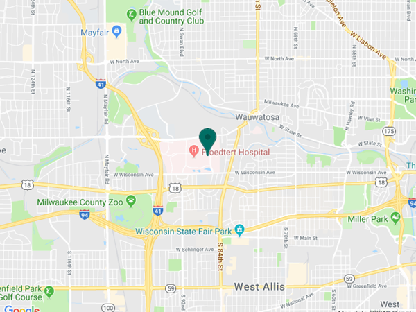
Eye Institute Google map location